Table of Contents
What You Need to Know | Causes and Types of Dog Skin Lumps and Bumps
How to Track Lump Growth and Changes | Diagnosis | Treatment | Ask a Question
Summary:
"Dog lumps on skin can happen for many causes from an insect bite that results in to an abscess where pus forms under the skin to lipomas (fatty tumors which are usually benign or not cancerous), skin tumors (malignant) or even cysts. This article discusses the dog skin tumors in particular. However, don't rush to judgment too soon or assume that a canine skin bump is cancerous, as many types are benign or not cancerous.
The diagnosis will depend on an understanding of if the lump has changed over time, rate of growth, the look and feel and if it is interfering with the quality of your dog's life. That said, 25% of dogs will develop cancer at some point in their life, making it imperative that any unusual scab, bump, lump or sore on the skin be investigated by a veterinarian. The most common type is a mast cell tumor with 20% of all cases (benign and malignant). There is no known way to prevent skin malignancies other than squamous cell cancer which is thought to be caused by sunlight.

The best thing you can do for a dog skin lump is to have a veterinarian take a look. Often the Vet will recommend watching the skin lesion and if it changes in appearance, then do a biopsy or use another diagnostic approach. If required, the tumor can be surgically removed or another type of treatment applied (radiation, chemotherapy)."
Causes and Types
Dog skin lumps are primarily due to fluid accumulation under the skin. There are multiple reasons why this might occur. In general, causes fall into two categories:
- Benign Dog Tumors (not cancer)
- Malignant Tumors (cancer)
Benign Dog Tumors (not cancer)
Characteristics of these lumps are no or slow growth and associated symptoms are usually indicative of other common reasons. It is possible that they will not change or take years to change. These growths can become large, although they are usually well defined and small.Common types of benign dog skin bumps include:
- Abscess: this is a cavity in the skin filled with pus, in general is accumulation of trapped dead white cells in walls as a result of bacterial infection. Abscesses are caused by pyogenic, i.e., pus forming bacterial or fungal infections, very rarely they can form at the sites of an injections. The abscess can be fluid-filled or firm. If the abscess becomes infected it can cause appetite loss and fever. Treatment involves draining the abscess and using antibiotics where needed.
- Acral Lick Dermatitis (neurodermatitis): Neurodermatitis is caused by licking the skin. It results in hair loss and possible a raised lesion, frequently on the leg. Treatment involves addressing the underlying cause of the licking such as stress or anxiety. An Elizabethan Collar can cut down on licking.
- Adenoma: these develop in the sebaceous glands of the hair follicles. There can be multiple adenomas on a dog, particularly in cocker spaniels and miniature poodles. (this is different from the cancerous adenocarcinoma that can also develop in the same location. A similar sounding perianal adenocarcinoma is also cancerous and develops in the tissue of the anus).
- Apocrine Sweat Gland Cyst: A type of cyst frequently found on the limbs, neck and head. It is round, smooth and has no hair. It can be firm or most contain a watery liquid. The veterinarian will do a biopsy to rule out cancer. Benign cysts do not need to be removed.
- Calcinosis Cutis (skin mineralization): In this condition hard papules and nodules form on areas such as the groin and back. It is caused by too many corticosteroids in the system. Treatment involves surgical removal or reduction in the use of steroids.
- Callus: A callus forms on the skin from pressure in bony areas or pressure points. Treatment involves use of a soft dog bed or the placement of padding in the treatment area.
- Canine Acne: Acne or papules forms (usually in younger dogs) when the hair follicles become inflamed due to a bacterial infection. Treatment involves the use of Benzoyl peroxide and/or antibiotics.
- Chiggers: A chigger is an insect that deposits larvae in the skin. It occurs on the ears, stomach or feet. A veterinarian will look at a sample under a microscope to diagnose the condition. The condition is treated with the insecticide Permethrin or Pyrethrin.
- Coccidioidomycosis (fungus): The skin fungus Coccidiodes immitis can cause nodules. It is found in the Southeastern United States. In addition to skin nodules common symptoms are weight loss, fever, difficulty breathing and infection where the bones press against the skin. Treatment is with the medications Itraconazole and Ketocanazole.
- Contact Dermatitis: When a dog is allergic to a substance in the environment it can cause skin blisters or red skim bumps. Other symptoms include hair loss and itch. Treatment involves identifying and removing the allergen from contact with the dog. Allergic or irritant contact dermatitis can be caused by medications, pet care products, or materials such as wool or rubber.
- Cutaneous horn: A cutaneous horn is a hornlike growth 1/2" to 2" with unknown cause. It can be a byproduct of either follicular cysts or cancer. Some dogs have an allergic reaction to the larva. Treatment involves surgical removal. Treatment involves opening the nodule and then removal of any larva.
- Cuterebra: This is a type of fly larva. A dog skin lump forms around the larva which has a tiny opening at the top that enables the larva to escape . Nodules are found on the neck and head.
- Diracunculiasis: This is a parasitic condition caused by a worm which causes a nodule to form. It is seen on the stomach, head, and legs. Treatment involves surgical removal.
- Elbow Calluses: Large dogs can get lumps and bumps where their body comes in constant contact with the ground. These lumps are usually calluses or hardened skin. These can be treated by applying petroleum jelly, lanolin or other dog safe softening products.
- Epidermal Cysts (infundibular cysts): These types of cysts are 2" nodules that have a thick sebaceous liquid inside. They are the result of a bodily reaction to skin cells. They are treated with surgery.
- Flea Allergy Dermatitis: Dogs that have a more intense reaction could develop skin papules, scales and crusts. Itching can lead to infection. The veterinarian will test the skin for fleas. Treatment includes antihistamines or steroids for inflammation and itch.
- Granuloma: A granuloma forms after an infection or exposure to an irritant such as a plant. It results in the formation of a skin nodule. Granulomas can have different sizes and can cause a skin ulcer, hair loss and skin infection. Treatment involves removal with surgery, medications to treat any infection and removal of any underlying reason for the problem such as plant or chemical exposure.
- Hematoma: A dog hematoma is a nodule that contains fluid. It is found in different shapes. It occurs when blood leaks out of a blood vessel. Typically found in dogs with pendulous ears and as the result of an ear infection.
- Histiocytoma: A tumor found in young dogs that results in a raised red nodule. It looks like a strawberry. Histiocytoma tumors are usually seen on the ears, head and legs. They often go away with no treatment, but can be removed with surgery.

- Histioplasmosis: It is a non-contagious skin fungal infection that can cause skin lesions. Other possible symptoms include respiratory distress and problem with the digestive system. The fungus may localize in bone marrow and eyes.
- Hives (urticaria): Hives are often a reaction to something in your dog's environment. Allergic reactions to bug bites or a medication can cause the skin to swell from 1/2 inch to 2 inches. You could try Benadryl in the dosage 1 mg per pound of body weight. If ineffective, see your veterinarian. Another name for hives are Wheals which tend to look like a circumscribed, circular, raised area of skin caused by swelling due to the retention of fluids in the skin tissues (edema). Wheals are seen at the site of positive skin test reactions when dogs are allergy tested. Hives can go away without treatment. If the hive is the result of an allergy than a veterinarian will prescribe corticosteroids, epinephrine or an antihistamine.
- Hookworm: The hookworm can cause red dog lumps on skin. The problem is on on the foot pads. a veterinarian will administer a hookworm treatment.
- Kerion: A kerion is a skin nodule that is related to a case of dog ringworm. The condition can cause skin ulcers on the dog's nose and ears. Treatment involves removing hair around the infected area and treatment with a medicated shampoo that contains itraconazole or ketoconazole.
- Leishmaniasis: Leishmaniasis is a blood parasite. It can cause ulceration on the ears and nose. Contagious to people. Cannot be cured with a poor prognosis.
- Lichenoid Dermatosis: Flat skin nodules that is covered with a thick surface. Usually clears with no treatment.
- Lipomas: Canine lipomas are fatty tissue just under the skin surface about the size of a large coin, but could grow to the size of a large ball. They are often seen in middle aged, overweight female or older dogs and tend to appear on the belly and upper legs. Lipomas are usually seen in Schnauzers, Labs, Dobermans and mixed breeds. It is rare for a lipoma to be cancerous. Sometimes they will develop at the top of a forelimb, making it difficult for the dog to run or walk. If this is the case it will need to be removed with a surgical procedure.
- Medication Skin Reaction: Dogs may react to the site of a drug such as cephalosporin, sulfonamides and penicillins. Other symptoms include swollen skin, crusts, skin ulceration, redness and itch. Treatment requires not using the drug that caused the problem.
- Nocardia: A nocardia is a bacterial skin infection that is found at the site of a dog puncture wound. Other symptoms include breathing problems. The prognosis for the condition is not good with treatment involving the use of antibiotics.
- Panniculitis: When the skin is injured, or a foreign object enters the skin it can cause a nodule to form or skin ulceration. Typically found on the limbs or head. Other symptoms include appetite loss. The veterinarian will drain the nodule and reduce inflammation with prednisone.
- Pelodera dermatitis: Pelodera dermatitis is a type of skin infection caused by a worm larvae. It occurs and areas of the dog's body that comes in contact with the ground. Other possible symptoms include skin redness, loss of hair, scales, crusts and the formation of papules. Can also cause skin itch. Treatment involves the use of an antibacterial shampoo and steroids for skin inflammation and itch.
- Phaeohyphomycosis: A dog fungal skin infection that results in the formation of a skin nodule or ulcer. A Veterinarian will drain the wound and due a skin biopsy. Treatment involves removal with surgery and anti-fungal skin medications.
- Pyoderma: Pyoderma is the formation of nodules or pustules in areas where there is skin trauma caused by the dog such as licking. It can also be the result of conditions such as seborrhea or skin allergies. Treatment includes removing hair in treatment area, addressing any bacterial infections with anti-biotics and keeping the dog from licking the treatment area such as with an Elizabethan collar.
- Pythosis: This is a mold that causes ulceration on dog skin. It is frequently seen on the base of the tail, head or legs. It can cause itch and digestive problems. A Veterinarian will do a biopsy, drain any nodules and remove them with surgery. Can be fatal to the dog.
- Ringworm: Ringworm in dogs is a fungal infection that results in loss of hair, pustules and vesicles. Other symptoms are skin itch. Ringworm is treated with topicals such as Miconazole, a lime sulfur dip or products that contain itraconazole or giseofulvin.
- Sarcoptic Mange: Mange is an parasitic infection caused by a mite. It causes skin papules, scaling and crusts. It is diagnosed via a skin scraping by your vet. Treatment is with dips and the insecticide ivermectin.
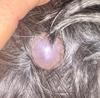
 Sebaceous Cyst on Dog Back
- Sebaceous cysts (Follicular Cyst): pea-sized single sacs filled with a thick liquid or cheesy substance. These dog lumps on skin are caused by an oil producing gland called the sebaceous gland. If the gland gets blocked it gets enlarged as small as a pen eraser and as big as 2 inches wide. If the cyst opens you will see a white pasty substance come out. Usual locations for sebaceous cysts are the trunk, neck and head. These cysts can be removed with surgery and can refill if punctured. To avoid skin reactions DO NOT SQUEEZE.
- Pimples/blackheads: Similar to people, dog skin can have the same types of problems. Like humans, you can rid your dog of pimples through cleaning clogged pores and the use of benzoyl peroxide. Another name for pimple is papule which is defined as a small solid rounded bump rising from the skin that is usually less than 1 centimeter in diameter (less than 3/8 inch across). Papules may open when scratched and become crusty and infected. Dermatologists (and other physicians) call any small solid circumscribed bump in the skin a papule, as opposed to a vesicle which contains fluid or a macule which is flat and even with the surrounding skin.
- Sporotrichosis: This is another type of fungal infection that occurs at the site of a skin puncture. It results in a raised dog skin lump (nodule). Treatment includes draining the nodule and use of itraconazole, ketoconazole or potassium iodide.
- Stings and Bites by Insects: Dogs can have an allergic reaction to an insect sting such as hornet, wasp or bee. The skin reaction can result in swelling and redness or itch. If skin ulcers form they will need to be trained and treated. The allergic reaction may require treatment with steroids.
Spider bites are called eosinophilic folliculitis and swell right after being bitten (also is caused by bites from caterpillars). These bites are seen on a dog's nose. Treatment is with wet dressings and corticosteroids.
Ticks can cause skin inflammation resulting in a nodule where the bite occurs. The nodule can persist for a few months. Treatment involves tick removal and a tick preventative. The nodule will heal without treatment. - Tail Gland Hyperplasia: In this condition a gland enlarges in the tail causing a dog skin lump. The sebaceous gland becomes larger, a problem seen in un-neutered dogs and in dogs that suffer from hypothyroidism.
- Zygomycosis: This is another disorder caused by a fungus. It results in skin nodules along with symptoms such as jaundice or vomiting.
- Warts: Warts are rough surfaced lumps that are hard, firm bumps that are often the result of a viral infection. They are seen in young and old dogs. Young dogs get warts from a viral infection that are seen around the mouth. Warts in younger dogs go away by themselves. They can also be removed with cryosurgery (freezing).
Malignant Lumps (Cancer)
These lumps may grow quickly and spread into the skin. These dog skin tumors can grow uncontrollably and metastasize (spread) to other parts of the body. The most common type of are mast cell tumors and occur 20% of the time (benign and malignant types)
- Epitheliotropic Lymphoma (rare): Lymphocyte cancer that can cause skin ulcers, oral ulcers and nodules, particularly in regions near to lymph-nodes. The disease can be primary in nature but also appears as secondary to whole-body, internal diseases such as canine malignant lymphoma. Many treatments for the condition have been tried, although none of it has proven to be completely effective. Treatment includes chemotherapy and surgery, which may improve the signs of disease but doesn't contribute in lengthening the life of dog.
- Fibroma (uncommon): Dog skin tumors that form in fibrous tissue that is held on with a stem like structure called a pedicle. Usually forms one nodule that is seen on the sides, groin or legs. These resembles to skin tags, which are usually distinctive, benign and appears as skin lumps on skin. Fibromas appears firm but rubbery, while other kinds can be soft and mushy. Usually the surgical removal is optional but complete surgical removal is recommended in case the fibromas appear to be growing and change appearance over time.
- Fibrosarcomas: This is the type of dog tumor seen at the skin location where there was a vaccination. It is a firm nodule that may become an inflamed skin lesion. Fibrosarcomas are an invasive and fast growing tumor and need to be surgically removed. The fibrosarcomas under skin appear as lump on skin, and in case multiple tumors are present, they usually occur in same area. If the tumor is large or very invasive, more area needs to be removed such as an entire leg. Radiation and chemotherapy are frequently used with surgery. Recurrence is common, 70% cases will see return in 1 year time. Therefore, follow-up radiation and chemotherapy is recommended after surgery, especially for the tumors which are inoperable or the ones which can't be removed completely. Fibrosarcomas appear fleshy and deep in muscles, thus complete surgical removal always remain questionable.
- Hemangiosarcoma: It is very invasive tumor that occurs on skin that was damaged by the sun. Causes a skin nodule that is black to red in color. Typically located on the abdomen or chest. Can turn into a skin ulcer.
- Hemangiopericytomas: canine skin tumors that form near blood vessels, thus spread in whole-body makes the prognosis quite poor.
- Hematoma: a cavity in the skin filled with blood. A Hematoma is usually caused by trauma or injury.
- Histiocytosis: This is a malignant tumor that can be found internally or as a skin disease. Diagnosis is with a fine needle aspirate. There is no effective treatment. Dog may have to undergo euthanasia when there is a poor prognosis.
- Lymphoma: This type of dog skin cancer results in skin nodules and ulcers. The veterinarian will confirm any diagnosis with a skin biopsy. Treatment involves surgery, radiation and chemotherapy.
- Mammary Cancer: Nodules found around the breasts. Treatment with surgery to remove the nodules. Half of all cases are cancerous.
- Mast cell tumor: These types of cells are essentially white blood cells that are used by the body to defend against outside elements that enter your dogs body. Primarily, mast cells have a function related to allergic reaction and release histamines. For example, an insect bite causes swelling on the skin because of these cells. If the normal mast cell undergoes a malignant change, a mast cell tumor may be produced. They can be malignant or benign and are found in the bone, cartilage, the skin and other tissues. The cells contain histamine which is a chemical released by the dogs immune system. Mast cell tumors are the most common type of skin tumors in dogs and can affect dogs of all ages and most breeds. The tumors, which aren't removed completely can spread to other parts of body.
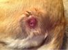
 Dog
Mast Cell Tumor Picture on Dog's Skin
Dog
Mast Cell Tumor Picture on Dog's Skin- Melanoma: A melanoma is a cancerous tumor frequently seen in older canines. Symptoms involve a single skin lump. A Vet will do a biopsy to confirm the diagnosis and then surgery to remove the nodule along with some surrounding skin to avoid any spread of cancerous cells.
- Neoplasia Nodule: a firm, solid cutaneous swelling up to 1 cm in diameter caused by infiltration of inflammatory or cells that are abnormally uncontrollably growing (neoplastic cells).
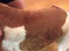
- Squamous Cell Carcinoma: These are flat scaly cells on the outer skin layer. Dogs that have white coats or lightly pigmented skin are predisposed. A contributing factor is sun exposure such as dog that frequently basks in the sun while lying on its back. Treatment involves removal surgically.
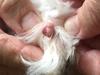
 Dog Lumps on Skin Pimple or Papule
Keeping Track
Janet Tobiassen Crosby, DVM in Your Guide to Veterinary Medicine has some excellent advice for keeping track of dog lumps on the skin. She recommends the following approach:
- Take a piece of wax paper and a felt marker.
- Lay the wax paper over the lump. Trace the outer edges of the lump.
- Date the wax paper.
- Repeat monthly (or shorter intervals, if your vet suggests) to monitor.
Canine Skin Bump or Lump Diagnosis
Your Veterinarian will look at the physical characteristics of dog lumps on the skin to determine a preliminary diagnosis. They will look at the location, duration, firmness, and size of the canine skin lump or bump.
According to Dr. Timothy Fan, your veterinarian will ask several questions about the dog lumps on skin or bumps:
- Has the lump appeared suddenly, or has it been there awhile?
- Has the lump stayed the same in consistency and appearance or has it changed recently?
- Does the lump seem to separate from underlying tissue or is it attached?
- Is there only one lump or have you found multiple dog lumps on skin?
- Are there changes in your dog's behavior such as eating less, losing weight, vomiting, diarrhea, or lethargy?
Common Signs of Dog Neoplasia (Tumor)
- Abnormal swelling that does not stop growing
- Sores that fail to heal
- Loss of weight
- No appetite
- Any bleeding from any part of the body
- Strong offensive smell or odor
- Trouble swallowing or eating
- Reluctance to exercise, low energy
- Acting lame or stiff frequently
- Respiratory issues, urination issues, defecation issues (trouble going to the bathroom)
To be sure of a diagnosis of dog lumps on skin, the veterinarian will take a sample by using a needle to remove some of lump for study in a laboratory (fine needle aspirate).
Treatment

Source: Washington State University
For Lipoma, your Veterinarian will probably watch and wait to see if the identified dog lumps on skin shrink or stop growing. If the lump interferes with your dog's quality of life or is growing, then surgery is used to remove the lump.
Warts in younger dogs will go away by themselves. In older dogs they will need to be removed with surgery if they are causing any kind of problem (bleeding, irritation).
If the dog lumps on skin or bump is malignant, then routine cancer therapy including surgery, radiation and chemotherapy are used. It can be difficult to determine the best treatment option. While cancerous conditions cannot be prevented, overall skin health may improve with homeopathic supplements such as Skin & Coat Tonic, which are formulated to support the systemic health of the skin and C-Caps which promotes cellular health and immune system function.
Prognosis
In general, a dog that has a dog skin growth removed surgically will be active in a few hours after the procedure and will not need a look recuperation period. If chemotherapy is used then the recuperation is longer. If the growth has been completely removed, then there is a good change the growth will not return, leading to an excellent prognosis. Prognosis in general refers is related to the size of the tumor and the ability of the surgeon to completely remove the growth. It mostly depends on the nature of tumor, whether benign or malignant.
Ask Our Vet A Question And We Will Answer It For Free
Ask Our Vet A Question About Your Dogs Skin Lumps or Condition. We Wil Answer It For Free!
Do you have a question for our veterinarian about dog skin problems?
Our editors will pick 1 question to answer each week. Please include your dog's age, breed, sex, medical history and if possible, a picture of the condition. Include important details such as changes in behavior, when the condition first appeared, medications, and any changes in your dog's grooming or dietary routine.
We will do our best to get back to you quickly (it depends on how many questions we receive each day). If you do require an immediate response we suggest using this online dog veterinary service that is available now.
Other Reader Dog Skin Related Questions and Vet Suggestions
Click below to see contributions from other visitors to this page...
Treating a Dog With a Lump on Head 
Reader Question: My dog is a 12-year-old rescue Lhasa apso, Radar is a male and has been fixed. Last year I found a few lumps under his skin and the Vet …
Is This Picture a Cancerous Lump on my Dog Not rated yet
My lab is 12 years old. This lump appeared this summer. Can this be safely removed?
Editor Suggestion - Dog Lump Removal
Hi,
It is impossible …
Understanding Causes of Dog Skin Lumps Not rated yet
Reader Question
My Bichon mix has this growth on her right front paw. She had been licking it for at least 2.5 months but it was smaller and has …
Dog Lipoma Not rated yet
My black lab'hound mix, 12 year old male has a bubble like growth on his forehead, started at least 5-6 months ago and has gotten larger to the size of …
Treating Dog Skin Bumps Above The Eye Not rated yet
Reader Question Hi there! My French Bulldog, Walter is nine months old and is an intact male. He is up to date on all his vaccinations and is a very …
Cause of Dog Skin Lumps on Sides and Tummy Not rated yet
Reader Question: What could these dog skin lumps be? I squeezed them and nothing came out. He has had them for about a week. Could they be related to my …
Brochures
References for Dog Lumps on Skin
- Washington State University
What to do if you find a growth on your pet - Sarah Probst
Information Specialist
University of Illinois
College of Veterinary Medicine - Recent Advances in Mast Cell Tumors
K.A. Selting
College of Veterinary Medicine, University of Missouri
Columbia, MO, USA - Brevitz, Betsy DVM
Hound Health Handbook - Understanding the Language of the Skin
Peter Hill, BVSc, PhD, DVD, DipACVD, MRCVS
The Royal (Dick) School of Veterinary Studies
The University of Edinburgh, Scotland - Merck Veterinary Manual (11th Edition)

